Technologies
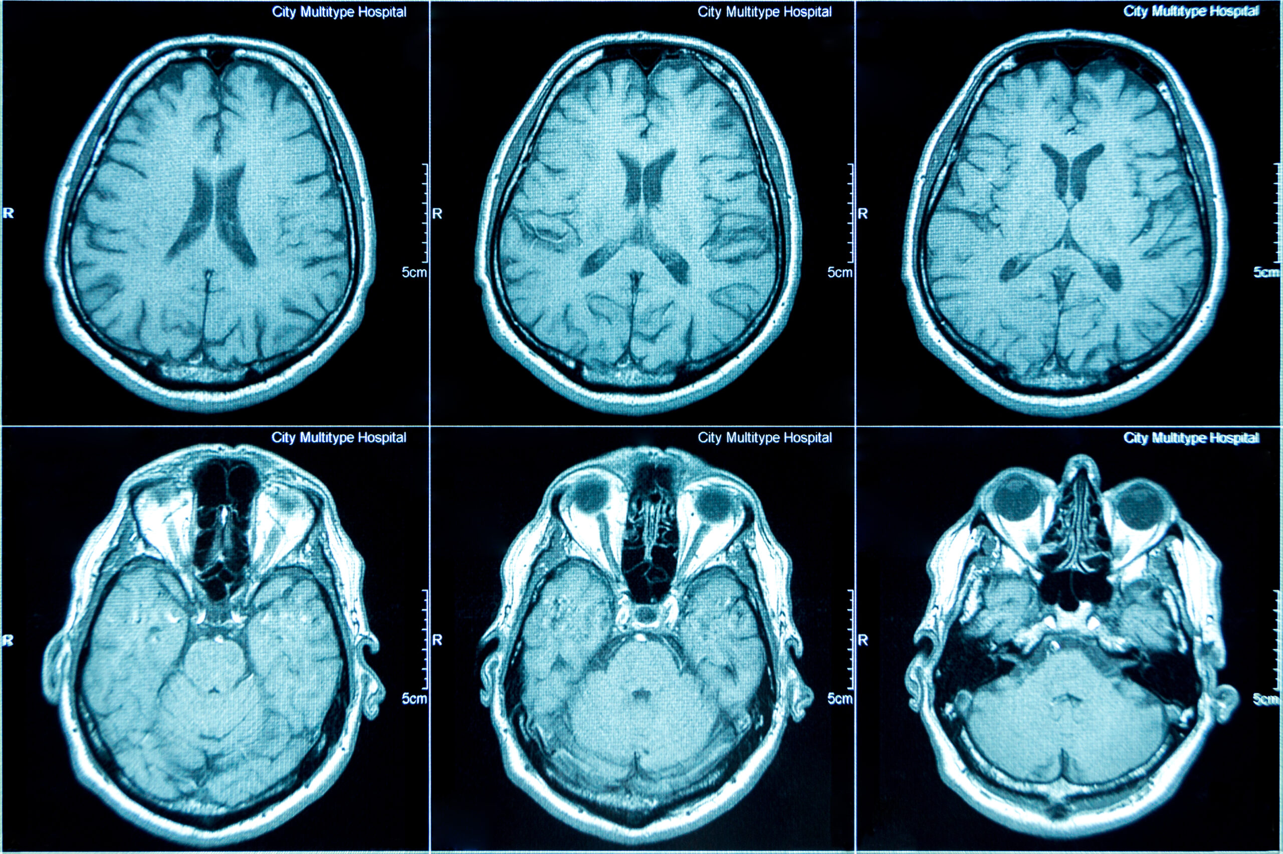
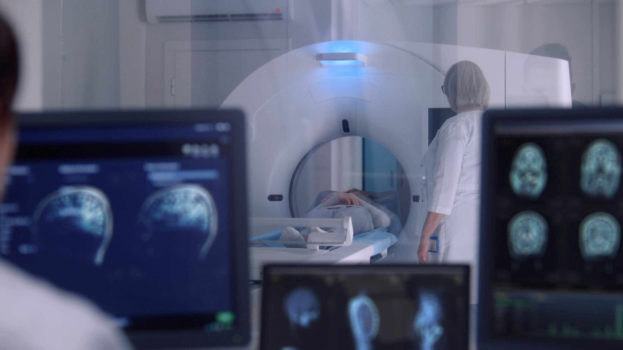
Positron Emission Tomography (PET)
Positron Emission Tomography (PET) is a powerful medical imaging technique used to visualize and quantify physiological processes within the body. Unlike structural imaging methods, such as Computed Tomography (CT) or Magnetic Resonance Imaging (MRI), PET provides functional information by detecting the distribution of biologically active molecules labeled with radioactive isotopes (tracers). After the tracer is administered—typically by injection—it accumulates in specific tissues, and its decay releases positrons. When these positrons interact with electrons in the body, they produce pairs of gamma photons that are detected by the scanner to reconstruct detailed images of metabolic activity, blood flow, or receptor binding.

Multiphoton Endoscopy (ME)
Multiphoton endoscopy is an advanced optical imaging method that enables high-resolution, three-dimensional visualization of living tissues deep within the body. It works by delivering ultrafast infrared laser pulses through a miniature endoscope, causing certain molecules within the tissue to emit light through a process called multiphoton excitation. This allows clinicians and researchers to image structures such as cells, collagen, and blood vessels in real time, without the need for dyes or tissue removal. Because the technique minimizes light scattering and tissue damage, it is especially valuable for examining delicate or hard-to-reach internal areas with high precision.
Image Co-registration
Multiscale image coregistration aligns images at multiple levels of resolution, beginning with coarse features and progressively refining to finer details. Artificial intelligence enhances this process by learning complex spatial relationships between images, enabling accurate alignment even across different modalities or timepoints. This approach ensures fast, robust, and precise image registration, making it ideal for applications like medical imaging, microscopy, and time-series analysis.